Deciphering ultrasound during pregnancy 7 weeks. Ultrasound in the seventh week of pregnancy. Conflicting emotional state
- What's happening
- Fetal development
- ultrasound
- When should you not sleep on your stomach?
- What you need to know
The 7th week of pregnancy is not the easiest for many. There can be no more doubts - pregnancy has come, this fact is confirmed. Now the daily life of the expectant mother begins to change rapidly every day. We will tell you more about what happens at this time with the baby and the woman in this article.

How many months is this?
7 weeks pregnant is the obstetric period. For convenience, doctors use a special calculation system - they count from the first day of the last menstruation. Thus, the real term differs from it by about 2 weeks, because in the first 14 days of the cycle it is impossible to get pregnant due to the immaturity of the egg. 7 weeks by obstetric standards is 5 weeks from conception.
The embryonic period is about five weeks if ovulation occurred on time, in the middle of the cycle.

This period may be slightly less if ovulation was late. 6-7 obstetric weeks, which will be discussed, are almost two obstetric or lunar months of pregnancy (they last 4 weeks each). In a more familiar calendar calculation of time, the second month of pregnancy began. It's been three weeks since the start of the delay. The baby is now not just not dividing cells, but a full-fledged embryo, and in his five weeks in his mother's womb he accomplished much.
embryo development
The baby is officially still considered an embryo, but in fact, a new stage for him begins this week - the neo-fetal period, which will last another 2 weeks. After 9 weeks, babies are officially called fetuses. These 14 days between weeks 7 and 9 are considered the transition period from the embryonic stage to the fetus. The most critical period in which no one makes predictions about the life of the child and the continuation of pregnancy is coming to an end.
From the seventh week, there are less and less threats to the development and life of the crumbs, his chances of surviving and being born into this world are increasing. Many doctors no longer call babies embryos and speak of them as fetuses.

Height and weight
The baby is growing, and so fast that its parameters change daily. All the energy, all the forces of the baby and the mother's body are now thrown precisely into growth. This week, at the very beginning, the growth of embryos averages about 2 centimeters, but by the end of the week it can reach 5 centimeters. The weight of the crumbs this week is less than a gram, the baby will begin to gain weight later.
To visualize what your baby looks like now, it is worth remembering what a medium-sized white bean looks like - this is what a child is now.
Nervous system
The largest part of the baby's body this week is the head. And this is not just because it is at this time that the brain undergoes the most intensive development. Five bubbles are formed in it - cavities that will correspond to each of the sections of the brain. The medulla "adds" in weight and volume, the brain is divided into two hemispheres. The neural tube is not yet closed, the formation of the nervous system is underway.
This week begins the development of nerve fibers that will connect the "control centers" - the central nervous system and all internal organs. How favorable the conditions in which the baby develops at the 7th obstetric week depends on how correctly its nervous system will form and work.
Some congenital chromosomal defects, if the fetus has them, do not allow the brain to form at this time, such babies die. At this time, a woman should remember that it should not be exposed to radiation, toxins, most medicines, since all this can totally and irreparably affect the formation of the child's brain.

Internal organs
This week, vision begins to form. The anterior cerebral bladder begins to bulge somewhat forward. It is he who will give rise to the optic nerves and retina. A woman should remember that at week 7, she should not be deficient in vitamin A and folic acid necessary for the proper development of the organs of vision and the nervous system of the crumbs.
The biggest changes at this time occur in the baby's digestive system. The intestine, so far the only one, is divided into main sections, which will soon be full-fledged esophagus, stomach and pharynx. The middle part of the intestine begins to go towards the umbilical cord. The back of the intestine becomes more complex these days, forming the urogenital sinus and rectum. The bile ducts appear, the appendix is laid, and the pancreas will begin to produce its first insulin this week.

The respiratory organs are represented only by the trachea. But the lungs and bronchi are already forming and growing. But a big "breakthrough" is planned in the cardiovascular system.
The heart becomes four-chambered, now it can pump blood throughout the baby's body. The network of blood vessels expands, grows, and large blood vessels develop.

Floor
At week 7, the so-called genital folds begin to form at the primary kidney. They are the rudiments of the sex glands. In place of the external genital organs, only the genital tubercle is formed so far, which looks completely identical in both boys and girls.
Sex is predetermined from the moment of conception. The chromosomes in the last pair matched either as XX, and then the little princess was destined to be born, or as XY, and then a boy would be born. However, there is currently no way to determine gender. The sex glands of the fetus do not yet produce sex hormones, the external genitalia have not formed.

Appearance
A small creature inside the mother's womb every day will more and more resemble a person. In the meantime, the baby looks like an alien. The rudiments of the eyes are set on the sides of the large head. Every day they will gradually "move out" to the center of the face - to where the formation of the rudiments of the nose has already begun.
This week, the inner ear is formed and the places where the auricles are destined to be are “marked” with pronounced dots with cartilaginous tissue. At week 7, the rudiments of milk teeth are laid and the jaws begin to form.
The spinal column is growing, this week the first cervical bend appears in the crumbs. The kid begins to move - so far only he can only unbend and bend like a small caterpillar. Spatulate prototypes of the hands and fingers appear on the handles, the nails are not yet formed.

The embryonic tail is shortened by about half, in two weeks it will disappear completely, turning into the coccyx familiar to each of us.
The legs of the fetus develop somewhat more slowly than the arms, until the feet are formed on them, but drop-shaped rudiments have already appeared. It's not the most important thing right now. All the forces of the baby and mother this week are directed to establishing a connection - uteroplacental blood flow is formed.
A very young placenta has a thickness of no more than 1 centimeter - it still has to develop for a long time before taking on all the functions of nourishing and protecting the baby.

What can the baby do?
At week 7, the baby masters an interesting movement - he learns to move his arms and trains on his own wrists. It is these flexions and extensions of the upper limbs that essentially represent the very first hand swings. By the end of the week, the movements will be more confident.

What can an ultrasound show?
Ultrasound scanning, so beloved by all pregnant women, is not among the recommended examinations at this time. If the well-being of a woman who is preparing to become a mother is good, nothing bothers her, does not hurt and does not cause concern, there is no need to do an ultrasound. Such a diagnosis can be recommended if there is a need to clarify whether there is a pregnancy at all, whether it is uterine, whether there is a threat of interruption and other complications.
Often, at week 7, ultrasound is done to expectant mothers whose pregnancy became possible after the use of assisted reproductive technologies, for example, after IVF or intrauterine insemination, to clarify whether the baby is viable, whether he is one or there are two or three of them.
If you suddenly have to visit an ultrasound room at week 7, then you should not expect that there will be a lot of information.

The doctor at this time does not determine the sex, does not measure individual parameters, but only specifies how many babies develop, fixes their viability (heartbeat, movements - sometimes), and also correlates the parameters of the fetal egg with the obstetric period. For the 7th obstetric week, the following dimensions are characteristic:
The average internal diameter of the fetal egg at 7 weeks of gestation:
The shape of the fetal egg will also tell a lot: even, oval or round is a variant of the norm, and a deformed egg with uneven fuzzy contours will certainly alert the diagnostician.

To clarify the term, the coccyx-parietal size of the fetus can be measured, although it is still so small that not every ultrasound machine is able to fix it. If you are lucky to do an ultrasound scan on a good scanner with high resolution, then the KTR for this period is within the following standards.
Coccyx-parietal size at 7 weeks of gestation:

The yolk sac, which has not yet "transferred" to the placenta its responsibilities for feeding the child, is now determined within 4-4.5 mm. The fetal heart rate is in the range of 129-146 beats per minute. Heart rate may differ from this range, but not exceed 180 beats per minute and not fall below 110.
On the monitor of an ultrasound scanner, a woman will not yet see her baby in all its glory. Most likely, the doctor will be able to show the expectant mother only a dark dot - fetal egg and a segment between two points in a dark cavity - KTR.

How does the expectant mother feel?
Everything that is happening in the body of the future mother is under the influence of hormonal changes. Immediately after fertilization, the production of the hormone progesterone increased, the task of which is to save the baby, prevent the uterus from straining and pushing the baby out of its cavity, and also not allowing maternal immunity to “deal with” the embryo, which is only half genetically related to the mother.
At the seventh week, the symptoms and signs of early pregnancy no longer leave any doubt. Under the influence of hormones, taste preferences have changed.
Progesterone increases appetite, because its "duties" also include the creation of fat reserves as an important energy reserve for the child and his mother.

Due to raging hormones, a woman becomes emotionally unstable. Her mood changes from amusement to tears in a matter of minutes. A woman feels unprotected, vulnerable, vulnerable, she may begin to perceive the films and photographs she has seen too sentimentally.
Since all the changes that occur with the baby and in the mother's body require huge energy costs, a woman may start to feel tired right after waking up in the morning. Dizziness, headaches, weakness, nausea are not excluded.
As uteroplacental blood flow begins to form, the concentration of the hCG hormone increases significantly, which leads to additional symptoms of malaise.

Hormonal changes can cause evening temperature rises to subfebrile values (above 37.0 degrees). Often women perceive this as symptoms of an incipient cold, but, apart from a slight chill, sensations of "burning cheeks" other signs of the disease are not observed. In the morning, the temperature does not leave a trace.
The size of the uterus has increased by one and a half times. Now it can be compared with the size of a goose egg or an orange. The growth of the uterus is not yet felt by the woman, she has not gone beyond the small pelvis, the feeling of heaviness in the lower abdomen does not yet arise.

Changes in a woman's body
Even if the daily work of a woman does not require her to concentrate her mental and physical strength, a decrease in working capacity at a given time is obvious. It is more difficult for a woman to concentrate, she is distracted, drowsy. This condition is considered completely normal for early gestation.
If the work of a woman is associated with high-precision production, then it makes sense to temporarily move to another position. If the work is associated with exposure to harmful substances, varnishes and paints, poisons, then the employer should be informed and switch to easier and safer work - such an opportunity for pregnant women is provided for by labor legislation.
Blood pressure this week may be below normal, and until a normal uteroplacental communication has been established, this is normal. Toxicosis, if it starts this week, manifests itself in all its glory.

Many women, speaking of toxicosis, mean nausea, but it is considered quite normal, and vomiting, especially repeated vomiting, are symptoms that require a doctor's consultation.
Severe repeated vomiting can be accompanied by dehydration, which is dangerous for both the mother and her baby. Therefore, toxicosis, accompanied by severe vomiting, which is repeated not only in the morning, but also during the day, necessarily needs medical correction, which can be provided to a woman in the gynecology department of the hospital where she will receive a referral.
A woman's skin cannot but respond to hormonal changes. This week, under the influence of hCG, it becomes looser, and therefore acne, rashes, dry fragments of the skin that itch and itch may appear. Dandruff may appear, although it was not there before. Some women complain of dryness in the vagina. All these are options for the reaction of the mother's body to hormonal changes.

The woman's chest becomes quite painful. It grows rapidly and increases in size due to the growth of glandular tissue, which is activated under the influence of progesterone. The nipples and mugs around them may darken, and not always evenly - sometimes pigmentation appears in spots with a more intense color. You should not be afraid of this. A comfortable, supportive bra can help relieve chest pain.
The intestines and digestive organs of a woman under the influence of progesterone may begin to work differently, and therefore, at week 7, increased gas formation, diarrhea, constipation, and heartburn may occur.
To minimize discomfort and symptoms, you should start eating right now so that the baby does not have a deficiency or excess of certain substances, vitamins, minerals, and the mother's body works more smoothly. Proper nutrition often helps to solve the problem of toxicosis at the same time.

pain
Small pulling pains on the sides of the uterus, as well as in the lumbar region, are not considered pathological. It's about the ligaments that hold the uterus. The main female reproductive organ is growing every minute, this dynamics does not stop, the ligaments are forced to adapt to this growth. This causes slight aching pain. Gradually, a woman gets used to them, they are no longer perceived as a cause for concern.
Other abdominal pains cannot be considered normal at week 7. First of all, cramping, severe pains are dangerous, in which the uterus comes into tone. Such a pain syndrome may indicate the threat of abortion, because at this time such risks are still very high.
Threat pains are similar to menstrual pains, only they are somewhat stronger.

If it pulls your lower back, your back hurts, it radiates to the rectum, the pain intensifies, it is important to see a doctor, because in most cases, with the timely intervention of a doctor, the pregnancy can be saved and delivered to the due date, then a completely healthy strong baby is born.
If your head hurts at week 7, this may be due to changes in blood pressure levels. It is important to monitor it daily, especially if the woman had a certain tendency to hypertension or hypotension before pregnancy. This should be reported to the doctor in the antenatal clinic, he will teach you how to measure pressure on both hands and keep a pregnancy diary. Pressure drops are fraught with the onset of preeclampsia - a condition that is extremely dangerous for the mother and fetus.

There is no cure for chest pain. They should simply be endured, because by the beginning of the second trimester they usually subside. Active growth of glandular tissue always occurs in the first third of pregnancy, as nature intended.
In general, various changes can occur with the expectant mother in this obstetric week, accompanied by pain. If previously a woman had any chronic diseases, right now they can worsen and make themselves felt with pain.

Allocations
Normal for this period are discharges that have a light color or milky color, odorless or with a slight sour smell. Any other discharge for a given period will be symptoms indicating that the pregnancy is proceeding with complications.
The most dangerous are spotting. Blood in any of its forms - from pink, creamy and brown discharge to open clean bleeding, can indicate a threat of interruption, spontaneous miscarriage, placental abruption or fetal egg.
If a bloody "daub" appears or a liquid, more abundant than just spotting, blood has gone, the woman should immediately call an "ambulance" and go to the hospital.
There are chances to save the child, but they will rapidly decrease if the woman continues to stay at home instead of seeking urgent medical help.

White thick yeast-like discharge, heterogeneous, with a pronounced sour smell, accompanied by itching and discomfort in the perineum and anus, may indicate the development of thrush. This disease is directly related to the changed hormonal background.
In this case, it is necessary to undergo treatment, the choice of the drug should be made by the doctor, after all, not all antifungal and antimicrobial agents are allowed for women in an “interesting position”.
Green, dark gray, brown, indefinite color discharge with an unpleasant odor, accompanied by itching, burning, is usually a manifestation of genital infections, inflammatory diseases of the genital organs. They must be treated taking into account the specific pathogen in order to avoid intrauterine infection of the fetus.

Possible problems
Since the 7th obstetric week is still a fairly short period, the likelihood of developing various complications remains high, despite the fact that the main threats to the life of the baby have passed.
What problems most expectant mothers face this week, we will tell in more detail.

Retrochorial hematoma
Every fourth pregnant woman can hear such a diagnosis. A hematoma forms in the space between the fetal membrane and the chorion if detachment occurs. The space is filled with blood, and this area is determined by ultrasound.
In most cases, a woman at week 7 learns about a hematoma not in the ultrasound diagnostic room, but by the first symptoms that make her see a doctor - by the appearance of a brown or pink “daub”, slight pulling pains. Despite the frightening symptoms, in most cases, pregnancy can be saved.

A hematoma may occur due to the fact that a woman overloads herself physically- lifts weights, professionally goes in for sports. Often, retrochorial hematoma is the result of a “jump” in hormonal balance, an echo of inflammatory diseases of the female reproductive system.
Pathology is often recorded in pregnant women who have not said goodbye to bad habits - smoking and alcohol, as well as in women who have problems with blood clotting and are exposed to severe stress.
Allocations with a hematoma are not an obligatory sign. Sometimes the pathology proceeds without symptoms at all. A brown “daub” with a confirmed pathology is a favorable sign, meaning that the blood from the hematoma is gradually moving away.

If a woman has scarlet blood, and the pain intensifies, it is likely that the hematoma has increased in size and is a great danger to the life of the child.
Favorable forecasts are usually about a hematoma, which does not exceed a quarter of the area of the fetal egg. If the size of the hemorrhage is larger, it all depends on the location and the exact size of the hematoma. Treatment is aimed at stopping bleeding, relaxing the uterine muscles with the help of antispasmodic drugs.

Non-developing pregnancy
A frozen or non-developing pregnancy is a pregnancy in which the fetus has stopped progressing and died, but continues to remain in the uterus. That's why pathology is sometimes called a failed miscarriage.
There are a lot of reasons why a baby stops developing, and not all of them have been thoroughly studied by medicine and science. Among the most probable experts call the chromosomal pathology of the fetus. If the baby at conception "inherited" extra chromosomes or they are missing, if he has a mutated gene that interferes with development and full growth, then the death of the child is inevitable.

In addition to genetic prerequisites, pregnancy can stop developing if the fetus is affected by toxins, certain drugs, poisons, radioactive radiation, and infections. Often, the true cause of the death of the crumbs at such an early date remains unidentified.
It is possible to detect a frozen pregnancy at week 7 mainly only on an ultrasound scan, which will show the absence of a fetal heartbeat and its significant lag behind the norm in size.
This week, many women go to the consultation for registration. Suspicion of a frozen pregnancy may appear at the obstetrician-gynecologist, who, at the first visit, conducts a manual examination of the uterus and cervix on the gynecological chair - the uterus will be smaller than the size due.

If the pregnancy stopped already 2-4 weeks ago, then right now obvious symptoms may appear - smearing brown discharge, indicating the beginning of the rejection of a dead fetal egg.
The pregnant woman should not be alerted by pain, the pain syndrome does not accompany the death of the fetus, and a sharp disappearance of pain and signs of pregnancy. If the chest hurt, it felt sick, and then suddenly all the symptoms disappeared, the chest stopped hurting, this may indicate that the baby “froze”. This can be determined by ultrasound diagnostics.

Ectopic pregnancy
An ectopic pregnancy is a pregnancy that takes place outside the natural place - the uterine cavity. A fertilized egg could be implanted 6-8 days after fertilization not in the uterine cavity, but to be fixed in the tube, in the cervix, outside the uterine cavity. Unfortunately, such a baby is doomed to death, but in addition to this, there is a great danger to the life of a woman.
If the grown fetus leads to a rupture of the tube, then the woman may begin massive internal bleeding, which can be fatal.

Some women who did not do an ultrasound before 7 weeks may be in the dark, because all the signs of a normal pregnancy will appear - nausea, dizziness, delayed menstruation, a change in smell, chest pain. At this time, a terrible truth may be revealed to a woman when registering in a antenatal clinic.
A manual examination on a gynecological chair will show the size of the uterus that does not correspond to the obstetric period, and therefore the woman will be urgently sent for an ultrasound scan, where it turns out that there is no fetal egg in the uterine cavity.
Depending on where exactly the embryo is entrenched, the symptoms of an ectopic pregnancy by week 7 may already manifest themselves. These are abdominal pains, fever, intestinal disorders, vomiting.

With the appearance of a sharp cutting pain, loss of consciousness, a rupture of the fallopian tube is likely, the woman should be taken to the hospital as soon as possible at any cost.
If a rupture has not occurred, and an ectopic pregnancy is detected during a routine ultrasound examination, then the chances of saving parts of the reproductive system for the woman are quite high. Laparoscopic surgery to remove the fetus from the tube allows you to subsequently become pregnant and carry the child quite safely. Cervical pregnancy is considered unfavorable, in which it is usually necessary to remove the entire uterus completely.

Anembryony
Approximately 15% of pregnant women are diagnosed with the absence of an embryo inside the fetal egg - anembryony. At earlier periods, such a diagnosis is often made erroneously, and the very next ultrasound shows that the baby is developing, his heart is beating. But at the 7th obstetric week, errors in diagnosis are practically excluded - the baby is already large enough, it is almost impossible not to see it and hear the heartbeat.
During anembryony, the fetal egg grows, its size increases, but there is no fetus in it. Experts believe that the pathology of the early embryonic period is to blame - when implantation took place, but the embryo inside the fetal egg died.
The most likely cause is called chromosomal genetic pathologies, the impact of negative factors - from a change in hormonal status to exposure to stress, medications, poor ecology, nicotine and alcohol, drugs, radiation. The reasons for the death of the embryo at the very initial stage of its development usually cannot be established.

If the absence of an embryo is detected this week, this usually causes serious psychological trauma to the woman, especially if the pregnancy is desired and the child is long-awaited. Such mothers usually have time to choose a name, look after a crib and a stroller, and resolve the issue of repairs in the nursery before the 7th week, so the news that there is no child can provoke a serious nervous breakdown in them. A woman may need qualified help from a psychiatrist or psychotherapist.
One consolation is that after anembryony, most couples become pregnant quite successfully and carry and give birth to a healthy baby without any problems. Cases of repeated anembryony are very rare.

Risk of miscarriage
This diagnosis in women's consultations sounds more often than all the others. According to medical statistics, about 70% of expectant mothers have a pregnancy with the threat of termination, most of them are diagnosed in the early stages. The reasons why a pregnancy is in danger of being interrupted at any time can be very diverse, but for the most part, doctors manage to cope with the problem and help a woman overcome a difficult period by maintaining a pregnancy.
Of course, no one can guarantee anything. Sometimes, despite the best efforts of doctors, a miscarriage still occurs. This usually happens if the child was initially doomed to death due to chromosomal abnormalities, total defects that prevent him from developing normally. Unfortunately, this can only be established posthumously - fetal tissue samples are subject to genetic analysis, which shows the presence of chromosomal abnormalities.

A miscarriage threatens women over the age of 35, in expectant mothers over 40, the probability of an unfavorable outcome is about 40%. At risk are women who have bad habits, work under conditions of constant stress, take medicines uncontrollably, abuse coffee or strong tea, expectant mothers who experience strong physical exertion.
Often miscarriages are habitual in nature - they repeat at about the same time. If the previous pregnancy ended in a miscarriage at 7 weeks, you should register as early as possible, to meet the “critical” week in a hospital under round-the-clock supervision of doctors.

The likelihood of a threat increases in women who have had abortions, including medical ones. After IVF, a miscarriage at 6-7 weeks and a little later ends approximately 25% of all pregnancies. And in women who have an immunological Rh-conflict with the fetus, the probability of miscarriage approaches 60%.
A threatening condition usually appears pulling pains or a feeling of "petrification" of the lower abdomen, abnormal discharge- bloody, watery, bloody.

Colds, SARS
Progesterone, which helps maintain pregnancy, weakens the immune system of the expectant mother. That is why in the early stages so often there are various viral and non-communicable diseases. If the flu or SARS started at week 7, it can be very dangerous for the baby, because the most important processes of brain formation are taking place in his body now, and high temperature increases the likelihood of miscarriage tenfold.
The situation is complicated by the fact that a woman cannot use the usual drugs for her treatment, which she used in similar cases before pregnancy - many drugs are now contraindicated for her, since they have a teratogenic effect (negatively affect the development of the fetus).

Seek medical attention at the first sign of a viral infection or flu. He will be able to suggest what drugs can be used, what folk methods of alleviating the condition a woman can use without the risk of harming the child.
An infectious disease at such a short time as the seventh week of pregnancy does not in all cases lead to irreparable consequences, Wrong treatment can be much more dangerous, to which a woman will resort without the permission and approval of the attending doctor.
For a cold, a woman can also take quite physiological manifestations of early pregnancy, for example, a runny nose. Nasal congestion in women at week 7 is often not due to hypothermia or a viral infection, but due to the action of progesterone on the mucous membranes. Under the action of the hormone, fluid begins to accumulate in the tissues, which leads to rhinitis.
If nasal congestion and difficulty breathing are not accompanied by additional symptoms of acute respiratory infections or SARS, there is nothing to worry about. Such a runny nose does not need treatment and does not harm the child.

Analyzes and examinations
Considering that most of the complications of pregnancy at such an early stage proceed without pronounced symptoms, it is advisable for a woman to get registered at the antenatal clinic as early as possible. It is better to do this at 5-6 weeks of pregnancy, but the seventh week is the best suited for a complete and detailed examination of the health of the expectant mother and baby.
When visiting a consultation, which can be scheduled this week, you should take your passport and medical insurance policy with you. If a woman has already visited an ultrasound on her own initiative, then you should take a doctor's opinion with you.

At the appointment, the doctor will draw up a detailed obstetric history of the woman, assess all risk factors, conduct a manual gynecological examination and issue directions for tests and examinations that are mandatory for registration. These analyzes include:
- general blood and urine tests;
- blood chemistry;
- blood test for infections, antibodies to TORCH infections;
- blood tests for HIV and syphilis, for hepatitis B and C;
- scraping from the cervix and a smear of vaginal secretion for microflora;
- colposcopy;
- coagulogram (blood test to determine clotting factors).

In addition, according to individual indications, other diagnostic studies may be prescribed for a woman. If there is a need to clarify the gestational age, an ultrasound is performed, if a multiple pregnancy is suspected, a blood test for hCG and ultrasound is done, tests for the content of hormones - progesterone, testosterone, thyroid hormones can be recommended.
When registering, a woman is recommended to visit other doctors who will give their opinions on the likelihood of complications from the internal organs and systems - a cardiologist, therapist, dentist, gastroenterologist, ophthalmologist, otolaryngologist.
If a woman has chronic diseases, the appropriate doctor should give written recommendations to the obstetrician-gynecologist about the specifics of managing this pregnancy, taking into account the disease.

To reduce the risks of complications in the seventh obstetric week, simple and uncomplicated safety measures that are desirable for all pregnant women will help.

Nutrition
If it's week 7, it's time to think about how to establish proper nutrition, which will help the baby get all the necessary nutrients, and it is easier for the mother to endure all the hardships of the "interesting situation". Whatever diet a woman adheres to before pregnancy, now it makes sense to make adjustments to it.
A woman should eat fractionally - 5-6 times a day. Portions should become smaller in volume, but more complete in composition and energy value.
You should completely abandon carbonated drinks, mayonnaise, ketchup, smoked meats, sausages and sausages. Salt intake is best reduced to 5 grams per day - this will help to avoid the likelihood of preeclampsia with edema.

It is also better to refuse factory sweets, chocolate, jam and preserves, preferring sweet fruits, natural juices.
The diet should contain fresh vegetables rich in fiber, which not only supply the mother's body with vitamins, but also contribute to the normalization of stools and prevent constipation.
Excluded fatty foods - lard, pork, lamb, large amounts of butter, margarine. It is desirable to minimize the consumption of bread, especially baked from rich or yeast dough. If a woman used to be fond of vegetarianism, now is the time to introduce meat into her diet - veal, beef, rabbit meat, poultry - chicken and turkey, because without animal protein the full development of the fetus is very difficult.

To withstand multiple meals, you should buy special food containers that will allow a woman to take food with her to work or school. Be sure to observe the drinking regime - drink at least 1.5 liters of clean water per day.
Sex
If a woman has no complications, pain, pathological discharge, if she is not at risk for miscarriage, then sex at week 7 is not only allowed, but also recommended.
A full-fledged intimate life has a beneficial effect on the psychological state of the expectant mother. The stomach has not yet grown and does not interfere, movements are not limited, it is no longer necessary to protect yourself.
All this makes a woman more liberated, she can get much more pleasure from making love than before. You should not worry about the child - the intimate life of the parents in no way harms his development, does not interfere with him.

Weight control
Starting this week, you should start controlling your weight. Many pregnant women acquire floor scales and at week 7 begin to keep pregnancy diaries, in which they indicate not only weight, but also basal temperature, as well as blood pressure.
There is no need to weigh yourself daily, it is enough to carry out one or two weighings (at the beginning and at the end of this obstetric week). You need to weigh yourself in the morning after going to the toilet, emptying the bladder and intestines, before breakfast.
A normal increase is now considered 100-300 grams per week.. If a woman has toxicosis, then there may even be some weight loss.

A pathological increase is considered to be an increase above 0.8-1 kilogram per week. This must be reported to the doctor in order to exclude the development of dropsy of pregnant women - preeclampsia.
Travel and flights
Travel, including by air, is not prohibited during the 7th week. The exceptions are cases of a clear threat of termination of pregnancy and miscarriages in the early stages in history, individual contraindications, which include, in particular, problems with blood vessels and the heart. Flights in this case are undesirable.

vitamins
You should not prescribe vitamins on your own. According to the results of a biochemical blood test, the doctor will be able to confidently judge what substances are missing in the body of the expectant mother. Based on this, one or another vitamin preparation is prescribed.
At week 7, the need for vitamin A increases, as the baby's organs of vision are formed. It is enough to introduce retinol-rich foods into the diet, for example, fresh carrot salad or cod liver.

Lifestyle
A woman at week 7 must definitely get enough sleep. It's time to forget about working on the night shift by asking management to change the expectant mother's work schedule.
If there are no contraindications, you can and should go in for swimming, walking in the fresh air. Excessive physical activity should now be eliminated, in any case, classes on power simulators in the gym are now clearly not shown.

The fifth week is the period when pregnancy can already be determined. The delay in menstruation is already two weeks, the state of health is changing, on the test, as a rule, the second strip is clearly visible. Is it dangerous to do an ultrasound at the 5th (7th obstetric) week of pregnancy and what it can tell about, let's talk.
At the fifth week of pregnancy, an ultrasound may not provide much information. During this period, a study is needed in order to confirm that the delay in menstruation, the change in well-being is not at all accidental.
 Many doctors believe that the expectant mother can do the first ultrasound examination later than at the fifth or sixth week of pregnancy (and it will already be more informative).
The only exceptions are those mothers who are expected to have abnormalities in the course of pregnancy.
Also, ultrasound at such times is necessary for those who have previously had an ectopic pregnancy in order to prevent a miscarriage and as one of the parts of prenatal genetic screening.
The condition of the mother and the development of the fetus during this period
At the 5th week of pregnancy, the neural tube has already formed in the child and the heart begins to beat, in addition, at this time the circulatory system, including the heart, is actively forming, and it will start working very soon.
All internal organs, bones and nervous system continue to form. The thyroid gland, pancreas and adrenal glands begin to develop. Moreover, they already secrete the first hormones, for example, thyroglobulin. From five weeks, the embryo can be called an embryo, as it enters the embryonic phase of development.

The size of the fetus is now 2-3 millimeters.
As for the mother's condition, there are also serious changes in it. During this period, all signs of pregnancy can make themselves felt:
- can pull the stomach;
- lethargy, constant fatigue;
- constant changes in mood;
- nausea;
- pain in the mammary glands;
- frequent urges in a small way;
- appetite changes;
- constant sleepiness;
- menstruation can be, but rather meager (their presence is not yet a bad symptom);
- sensitivity to odors.
But the absence of any signs and changes in well-being in the fifth week of pregnancy is the norm.
Important! During this period, the risk of miscarriage is high, in addition, the nervous system of the fetus is being formed, which means that mothers should not be nervous.
What will the ultrasound show?
At the fifth week, the ultrasonologist already perfectly sees that the uterus has increased. But this increase may not yet be completely uniform: for example, only the wall on which the vial with the embryo is fixed can increase and become convex.
The fetal bladder is already visible perfectly, its dimensions are about a centimeter. If you enlarge the image of the fetal bladder, then you can see the fetus itself and the yolk sac. A good specialist is able to recognize on ultrasound even the beating of the only emerging fetal heart.
But in the ovary, from which the egg came out, which was fertilized, you can see the corpus luteum. This is another confirmation that the pregnancy has come.

At this time, the configuration of the fetus already allows you to make assumptions about where his head will be located, and where - the lower limbs. By the way, their rudiments have also already appeared, but they are in the form of bubbles. Usually, a five-week-old fetus is actively moving, which allows the specialist to draw a conclusion about his health and proper development.
But ultrasound examination does not allow to consider anomalies in the structure of the fetus. But it can consider detachment of the amniotic sac, ectopic pregnancy or uterine hypertonicity.
During ultrasound, the specialist must measure and record the coccyx-parietal dimensions of the fetus, as well as its position in the uterine cavity, the condition of the corpus luteum, ovaries and myometrium. All these data will allow you to calculate the size of the baby and the approximate date of his birth.
Important! If, at a period of five weeks, the embryo is not visible in the fetal bladder and the doctor suspects a miscarriage, but you feel fine (no pain and bloody discharge), do not rush to do a curettage. Check for an ultrasound in another ten days (at least a week later).
Also, for a period of 5 weeks, you can already see whether the expectant mother is expecting one baby or she will have twins. The difference is that there will be two fertilized eggs, not one.
How do they do it?
Ultrasound during this period can be done both with the help of an abdominal and transvaginal sensor.

In the first case, the uterus and appendages are examined through the abdominal wall, in the second case, using a thin and long probe that is inserted into the vagina.
During the examination, the mother is shown the embryo and measurements of the fetal egg are taken.
After that, an examination protocol is made, which is given to the expectant mother.
Study preparation
It is quite simple:
- Do not forget to sign up and carry out the necessary hygiene procedures before the examination.
- A couple of hours before the ultrasound, drink gradually one and a half liters of water. You can’t go to the toilet yet, because a filled bladder is needed for the study.
- Also, do not forget about a diaper, shoe covers and a towel.
- Write down all your questions to the doctor (usually half of them are consistently forgotten in the office).
What can be seen in the photo at 5 (7 obstetric) weeks?
As mentioned above, sometimes an ultrasound may not show anything. If the hCG level is normal at the same time, and rapid tests consistently show pregnancy, you should not worry: an ectopic pregnancy at such times can already be seen. In the photo during this period, it is also often difficult to see something.

An ultrasound photo at this time may be blurry, and the embryo itself on it resembles a shrimp or a small cylinder in shape. If the picture and the apparatus are of high quality, then it will be possible to see the tail of the embryo and its head, as well as the rudiments of arms and legs, and sometimes even the oral and nasal fissures, eyes, fingers and auricles. As for the uterus, it should resemble an egg in its shape.
Twins at 5 weeks pregnant: photo

Norms of ultrasound examination
Having received the ultrasound protocol in hand, the expectant mother needs to contact her gynecologist to decipher the results.
For a period of 5 they should be as follows:
- embryo size - from 2 to 3 mm;
- the size of the yolk sac is 4-5 mm (if it is more than 6 mm, this may indicate a pathology);
- the size of the fetal egg is about one centimeter;
- heart rate - from 70 to 100 in 60 seconds.
There can be no other indicators for such a period yet.
Contraindications
In itself, an examination at such an early date can be called a conditional contraindication. More precisely, doctors do not advise doing this earlier: you won’t see much, it’s even impossible to determine gender, pathologies make themselves felt only at a later date.
About the exceptions in which ultrasound at such a time will be needed, it is written above.

It is difficult to say whether ultrasound will harm the embryo, because there is no evidence of harm, nor their refutation. So, if possible, sign up for the first one and do not disturb the growing fetus without any special need: 4-5 examinations in nine months will be enough.
Where can it be done and cost?
Ultrasound during pregnancy is done in all hospitals (both private and public) and perinatal centers. It is better to choose those that are equipped with the most modern and sensitive technology. Cost - from 3000-4000 thousand rubles per session.
Conclusion
Five weeks of pregnancy is not the best time for an ultrasound examination, and doctors do not recommend doing this examination. The only thing that ultrasound can tell exactly at a period of five weeks is whether there is a pregnancy or not. It will be appropriate only if the mother had problems with pregnancy before.
7 weeks of pregnancy is a wonderful period when some expectant mothers only find out about their piquant position by morning sickness, heightened sense of smell, irritability, and the absence of menstrual days. This is an important time when the gynecologist is obliged to register a woman, give a referral to narrow specialists and send her to take tests.
general characteristics
7 weeks of pregnancy is five weeks from the date of conception, or the completion of the second month (to be precise, one month is three weeks). Until the sixth week, a pregnant woman is not registered in any antenatal clinic. It is believed that spontaneous abortion before this period is natural selection. But from this period, a woman will not only be registered with a gynecologist, but will also be rushed to take tests before the twelfth week in order to receive additional social benefits for pregnant women.
A woman will have to prepare to bypass a huge list of doctors, but you should not be afraid of this. Since pregnant women will also be enrolled in the queue, the examination may drag on for a month or two (so you will smoothly move from narrow specialists to ultrasound, where you will receive a photo of the fetus). 7 weeks of pregnancy involves the delivery of the following tests:
- general examination of blood, urine;
- determination of blood group, sugar level, Rh factor;
- test for the detection of HIV, chlamydia, syphilis.
Be sure the gynecologist will refer the pregnant woman to a therapist, cardiologist, ophthalmologist, dentist. If a woman has health problems, then she will need to be observed by narrow specialists. Do not forget to remind the gynecologist about free vitamins for pregnant women. In many polyclinics, doctors simply do not issue prescriptions, hiding behind the financial “holes” of the polyclinic. Then immediately contact the head or the Ministry of Health of the region / city.
7th week of pregnancy: what happens to the female body?
The expectant mother feels the effect of hormonal surges with renewed vigor. Nausea, fatigue, drowsiness, vomiting, lethargy, lack of appetite, dizziness, weakness - these are a small number of manifestations of early toxicosis. This is due to the intensive development of the fetus, as well as the production of a huge amount of hormones, in particular, human chorionic gonadotropin.
To make yourself feel better, listen to your body. Feel apathy, fatigue, drowsiness - then drop everything and rest. But such recommendations are suitable for housewives. The following tips will help working women relieve malaise:
- in the morning, eat a green apple or drink a glass of cold water without getting out of bed;
- if you feel hungry, eat often, but in small portions;
- chew on nuts, crackers, or raisins if you feel nauseous;
- Suck on a lemon wedge or a sugar-free mint to avoid vomiting.
Try to eat those foods that the body accepts and requires at 7 weeks of pregnancy. Signs of toxicosis will soon pass, appetite will be restored, then the pregnant woman will return to her usual menu. In the meantime, the embryo receives the necessary nutrients from the mother, as well as the vitamin complex prescribed by the gynecologist.
How a woman feels at seven weeks
Due to hormonal intensive restructuring, a woman’s mood will also change, tearfulness, resentment, and irritability will appear. All this will pass immediately, as soon as the hormonal background stabilizes. In the morning, pressure may drop or rise, but it is better to report this to the doctor immediately. Hypertension, low hemoglobin may indicate the onset of serious illness in the early stages (meaning the 7th week of pregnancy).
What happens to the mother's body? A woman may experience breast tenderness due to their enlargement. In the second trimester, these discomfort will pass. Halos along with nipples, the middle line on the abdomen darken. Due to hormones, age spots, acne appear on the skin, and the number of moles increases. If a woman has problems with the thyroid gland, it is better to consult an endocrinologist. If the cause of the appearance of age spots is associated with hormones, then by the end of pregnancy or after childbirth, they will disappear.
In a pregnant woman, the total blood flow increases. The uterus begins to put pressure on the bladder, so the woman suffers from frequent urination. For uninterrupted sleep, try not to drink liquids at night, and also tilt your body forward while urinating to empty your bladder completely. The belly at the 7th week of pregnancy is invisible, but many mothers begin to eat for two, gaining weight intensively.

Meals for this period
Not only the full development of the fetus, the well-being of the mother, but also the normal course of pregnancy depends on nutrition. The first trimester includes five meals a day. This does not mean that every meal consists of three courses. The very last dinner can be at nine in the evening and consist of a glass of kefir. Remember that normal weekly weight gain is a maximum of three hundred and fifty grams.
By this time, the diet of a pregnant woman should include the following products that affect the development of the fetus (we will not paint by week, we will list only the daily rate):
- Two hundred grams of boiled meat.
- Two hundred grams of fish.
- One boiled egg.
- One hundred grams of cottage cheese.
- Fifty grams of cheese.
- Half a liter of milk or kefir.
- Six hundred grams of raw vegetables.
- Thirty grams of unrefined sunflower oil.
- Raw fruits or dried fruits.
- Black bread with bran.
- Buckwheat / oatmeal / barley cereals.
Please note that meat and fish alternate in a ratio of 5 to 2. Buy non-frozen foods.
What is unacceptable to do pregnant by the first trimester?
7 weeks pregnant is a tough time. The fetus has not yet fully consolidated, has not adapted to the mother's body, and therefore careless actions can lead to the threat of miscarriage. What can not be done at this stage of pregnancy?

Remember that heaviness, shock, radiation can lead to a threatened miscarriage. You can not do at this time what you did not do before pregnancy. That is, if you have driving experience, it gives you pleasure, then you can continue to use this type of transport. But if you are a beginner, you will only experience stress, tension, which is harmful to the development of the fetus. The same applies to air travel. If you have never flown and do not know your body's reaction to flying, then you should not experiment.
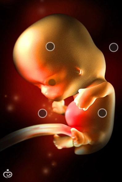
Fetal development by week: 49 days
Another week - and the embryo will take on a human form, but for now it looks like a tadpole. The head is almost proportional in size to the body, a tail is formed in the pelvic region. His height is approaching two and a half centimeters, and his weight is approximately nine grams. Development is progressing rapidly:
- The jaw, mouth, tongue, face are formed.
- Cartilage is replaced by bone tissue. Hands and feet with rudiments of fingers are visible. However, by seven weeks, a clear differentiation of the joints of the extremities is not visible on ultrasound.
- The auricles, nasal fossa, eyelids, pupils are formed, but the eyes are still tightly closed.
- In a nine-gram fetus, the genital organs are laid in the form of tubercles. In girls and boys, they have different forms, but it is not yet possible to determine the exact gender even by ultrasound.
Consequently, a child at the 7th week of pregnancy is comparable in size to a grain of wheat; by this time, the mother's belly is still invisible to others. It is possible to see your baby for the first time with the help of ultrasound from the eleventh week. Previously, this cannot be done, and it is not necessary, unless the pregnant woman is on conservation because of the threat of miscarriage.

Internal development of the embryo
If you resort to the results of ultrasound diagnostics, you can get more accurate information about the development of the fetus.
- According to the heart rate, there are up to one hundred and sixty beats per minute.
- The kidneys continue to develop, they are already divided into three parts, and in another week they will begin to function (discharge urine into the amniotic fluid).
- The digestive tract develops.
- The development of the placenta continues, which will form by the thirteenth week. This is an important organ that allows you to receive the necessary substances from the mother's body, as well as remove the waste products of the fetus. The umbilical cord of the fetus is attached to the uterus, so the right umbilical vein disappears. From this period, the fetus receives in full those substances that the pregnant woman consumes and inhales.
- At 7 weeks of pregnancy, the thyroid gland is actively formed, which is necessary for the production of hormones.
- The cerebellum develops, its work can be tracked by purposeful movements of the fetus. The brain also continues to form. According to ultrasound diagnostics, up to a hundred nerve cells are formed per minute.
What complications can arise during this period?
Pregnancy is different for every mother. Some may develop chronic diseases, others have chronic injuries, others, on the contrary, are cured of many sores and feel wonderful as never before. The reason lies in the hormonal background of a woman.

According to most pregnant women, the following troubles may appear by the beginning of pregnancy:
- Constipation, diarrhea. Change in stool is the body's normal response to the action of hormones at 7 weeks pregnant. The size of the fetus does not yet exert enormous pressure on the intestines, so you just need to follow a diet, drink more fluids, eat bran and fiber. Taking laxative pills, diarrhea medicines without a doctor's prescription is dangerous for the fetus.
- Heartburn. There is an unpleasant sensation due to malnutrition and hormonal failure. Requires a fractional balanced diet. Eliminate foods that irritate the gastrointestinal tract from your diet. Don't go to bed immediately after eating. For medications, it is better to consult a gynecologist.
Diseases that negatively affect intrauterine development

Dangers of 7 weeks of pregnancy
The greatest danger by this time is caused by an ectopic pregnancy, miscarriage (spontaneous abortion), missed abortion - only ultrasound can determine the initial symptoms. 7 weeks of pregnancy, according to expectant mothers, is the most dangerous period for the life of the fetus. It is important to avoid the causes that can cause this or that complication.

What should you do to keep your pregnancy going? The size of the fetus, its weight are indicators of intrauterine development, so do not miss an ultrasound scan, attend scheduled gynecological examinations, be sure to take tests. Watch your diet, do not undergo fluorography, try to avoid travel, especially air travel, avoid stressful situations, emotional and physical overload, work with a computer, chemical and radioactive substances, falls and blows to the stomach. If there are health problems initially, tell the gynecologist about them in order to avoid various complications in the fetus. Most importantly, listen to your body and intuition.
However, it is worth noting that each body reacts differently to the course of pregnancy. Someone at 7 weeks of pregnancy feels all the "charms" of toxicosis, and someone feels great throughout the entire period.
Already at 7 weeks there is an aversion to certain smells. It can be anything - from the aroma of fried potatoes to the perfume of her husband. During this period, certain eating habits may also appear. Someone is drawn to salty, and someone cannot spend a day without chocolate or cake.
It is worth remembering that it is during this period that the fetus is actively developing, taking all the nutrients from the woman's body, so it is very important to establish a varied diet with the inclusion of vitamin and mineral complexes.
Allocations in the seventh week
Transparent, odorless discharge in the seventh week is considered the norm, even if their volume has increased dramatically. During pregnancy, the blood flow in the genitals increases, the microflora and the structure of the vaginal mucosa change, and therefore the discharge becomes more abundant.
Alertness should be caused by yellow, green or brown discharge, which has an unpleasant, specific odor. This signals that the body is not all right. Especially dangerous at this time is the appearance of blood in the secreted mucus, which should not be at week 7. In this case, it is urgent to contact a gynecologist, since spotting indicates a threat of miscarriage.
Alertness should be caused by yellow, green or brown discharge, which has an unpleasant, specific odor.
If the secretion has become abundant and thick, resembling a curd mass, this is also a direct reason to see a doctor. Curdled discharge with a pungent odor indicates the development of pathogenic microflora, in particular a fungal infection. Most often, candidiasis develops against the background of a decrease in immunity and, if left untreated, closer to childbirth, it becomes complicated.Sex at 7 weeks pregnant
Experts note that sex during a normal pregnancy (if there are no medical contraindications) is important for a woman both physically and psychologically. Sexual intercourse stimulates blood circulation, removes pulling pains, causes positive emotions. In addition, the stomach at the 7th week of pregnancy does not yet prevent you from enjoying your sex life to the fullest.
Pulls the lower abdomen
At the seventh obstetric week of pregnancy, the expectant mother may experience occasional unpleasant pain in the lumbar region and abdomen. This is explained by the fact that the woman's uterus grows, slightly increasing the belly. These symptoms should not cause panic or active concern.
However, there is some possibility of abortion. Therefore, the expectant mother should be alerted if the painful discomfort is shifted to the lower abdomen. This situation requires expert advice.
Increased uterine tone and possible miscarriage can be avoided by following a number of simple rules. Give up shoes with high heels, make sure that your physical activity is moderate, avoid stress and strong emotional experiences, try not to take hot baths.
Embryo at 7 weeks of gestation and its development
At the term of the seventh obstetric week of pregnancy, as well as the entire first trimester of bearing a child, the embryo develops and forms at a "cosmic" speed. All organs and tissues grow so rapidly. The face of the future heir or heiress becomes more formalized and distinct. The shoulders of the unborn child are indicated. The embryo straightens, stretches. Movement of the upper and lower limbs becomes possible. This is from an external point of view. But what happens to the internal development of the bladder?
The whole complex of internal organs of the future person is also rapidly and actively developing. New cells in all organs and tissues grow every second at a tremendous speed.
The embryonic heart is already labeled as having four chambers. Very soon it will be able to take over the duties of blood supply to the entire small organism.
Large blood vessels and arteries appear. The brain of an embryo grows by leaps and bounds. Its division by structure into hemispheres, departments and zones is already being formalized.
Receive letters about the development of the baby and the condition of the mother once a week.
The seventh obstetric week of pregnancy is that milestone in the development of the embryo, when we can talk about the completion of the formation of the umbilical cord, which will be the "bridge" between the mother's body and the bladder for the next eight months. The umbilical cord begins to fully realize its main function - the transfer of nutrients to an actively developing baby.
The placenta - the environment between the mother and the unborn baby - becomes denser every day and continues to actively develop. A cork appears and takes shape, which will come off before childbirth, and for the next eight months it will protect and protect the fetus. The cork is formed by the glands of the uterine mucosa.
At this time, nature makes its choice of the gender of the unborn child - the formation of the sexual characteristics of the embryo takes place.
Now the length of the embryo is thirteen millimeters and its weight is approximately 1.7 g.
How does a woman's body change
Externally, the body of the expectant mother during this period practically does not change, however, some changes can still be noted. It is clear that the belly does not yet grow in this period, since the fetus is only being formed at the 7th week of pregnancy. But the chest, on the contrary, can increase by 1-2 sizes. This is explained by the preparation of the mammary glands for feeding.
An alarming symptom may be the presence of edema in various parts of the body. The presence of obvious swelling of the legs, fingers or face indicates that the pregnant woman urgently needs medical assistance and, if necessary, undergoing an additional examination.
Pregnancy is a process in which a baby develops from two tiny parental cells. The development of the fetus by week of pregnancy is a fascinating story about what exactly happens in each week of pregnancy, how the weight and height of the fetus changes, what feelings a mother has as the pregnancy progresses. In the article we will talk about what interests every expectant mother: when the baby begins to hear her speech, when and how the weight of the fetus changes, when you can take a photo of the fetus with ultrasound, what causes mother's feelings during pregnancy and much more.
First and second weeks of pregnancy: baby? Which child?
photo: 1 week pregnantAt the time of the appearance of the embryo, the gestational age is already 2 weeks. Why? Let's decide what we will consider the term from. There are concepts of embryonic and obstetric period. Embryonic gestational age - the true period from the moment of conception. Obstetric period - from the first day of the last menstruation. The obstetric period is on average 2 weeks longer than the embryonic one. During an ultrasound, in the card of a pregnant woman, on the sick leave, the obstetric period will always be indicated by the date of the last menstruation. But from the third week of pregnancy, the development of the fetus actually begins. Below you will find a description of each week of pregnancy: how the fetus develops, what happens to the uterus, how the feelings of the expectant mother change.
3rd week of pregnancy: parents meeting
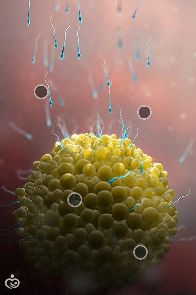
At the end of the second and beginning of the third week (on average, on the 14th day of the cycle), ovulation occurs. At this moment, the woman's egg leaves the ovary into the fallopian tube and where it meets with the sperm in the next day. Of the 75-900 million spermatozoa that enter the vagina, less than a thousand reach the cervical canal. And only one will penetrate the egg.
The spermatozoon and the egg cell carry half the set of chromosomes of the future person. As a result of their fusion, the first cell of a new organism with a complete chromosome set is formed - a zygote. Chromosomes determine the sex of the baby, the color of his eyes, and even the character. The zygote begins to divide and move to the uterine cavity. The journey to the uterus will take approximately 5 days, by which time the embryo will consist of approximately 100 cells. The next stage is implantation - the introduction of the embryo into the wall of the uterus.
4th week of pregnancy

The ball of cells is officially called an embryo. The size of the fetus at this time is like a poppy seed, approximately 1.5 mm.
At the end of this week, the expectant mother notices that the expected period does not begin. At this time, a woman may feel drowsiness, weakness, increased sensitivity of the mammary glands, mood swings. A pregnancy test shows a positive result. The test determines the hCG hormone, which begins to be produced after implantation.
The embryonic period lasts up to 12 weeks. There is a laying of the axial organs and tissues of the baby. A yolk sac is formed with a supply of nutrients, an amniotic sac, from these extra-embryonic organs, the fetal membranes and the chorion, the future placenta, subsequently develop. Below we will analyze what happens in the embryonic period every week, how the height and weight of the fetus changes, and what sensations a woman expects.
5th week of pregnancy

The embryo consists of three layers - the outer ectoderm, from which the ears, eyes, inner ear, connective tissue will form; endoderm, from which the intestines, bladder and lungs will develop; and mesoderm - the basis for the cardiovascular system, bones, muscles, kidneys, reproductive organs.
In the embryo, the anterior and posterior poles are determined - the future head and legs. The body of the embryo is laid along the axis of symmetry - the chord. All organs will be symmetrical. Some are paired, for example, kidneys. Others grow from symmetrical primordia, such as the heart and liver.
At the 5th week of pregnancy, with an hCG level of 500-1000 IU / l, it is possible to determine a fetal egg in size from 2 mm, this is the size of a sesame seed. Each woman experiences this period differently, but most experience nausea, drowsiness, intolerance to odors - signs of toxicosis.
6th week of pregnancy

Now the baby is no larger than lentils, at the beginning of the week 3 mm, and by the end - 6-7 mm. The embryo is somewhat similar to a fish and so far has little resemblance to a person. The rudiments of arms and legs appear. When the brushes appear, the legs will still be in the form of rudiments. The hemispheres of the brain are formed. A small heart pulsates, it is divided into sections.
From the villi of the chorion, the future placenta is laid, the vessels actively grow through which the exchange of blood takes place, and, accordingly, everything necessary for the unborn child between mother and baby.
At this time, the phenomenon of toxicosis may increase, severe weakness and vomiting may appear. It is important during these weeks of pregnancy to drink enough water.
7th week of pregnancy

An embryo about the size of a blueberry, height 8-11 mm, weight up to 1 g. There are hints of the future nose, eyes, ears and mouth. There is a fantastic growth rate of the brain - 100,000 cells per minute! Interdigital spaces have already appeared on the handles, but the fingers have not yet been separated. The umbilical cord and the uteroplacental circulation system are formed: the baby's breathing and nutrition comes from the mother's blood.
It is at this time that many expectant mothers often come for the first ultrasound during pregnancy. At 7-8 weeks with KTP (coccyx-parietal size) 10-15 mm. On ultrasound, a heartbeat with a frequency of 100 to 190 beats per minute is determined, which is much more than in an adult. At this time, the first photo of the gallery of fetal development is taken by week. Without instructions from a doctor and do not understand where to look. Later it will be clearer, especially on a three-dimensional ultrasound.
So far, the mother does not notice an increase in the abdomen, and the gynecologist can already say about the increase in the uterus. A woman has increased urination, which is associated with an increase in the volume of fluid in the body.
8th week of pregnancy
The baby is the size of a bean, from 15 to 40 mm, and weighs about 5 grams. Over the past two weeks, it has grown 4 times! The contours of the face continue to develop, they become more elegant, the upper lip, the tip of the nose stand out, the formation of the eyelids begins.
At the 8th week of pregnancy, ossification of the bones begins - arms, legs, skull. The structuring of the gastrointestinal tract, heart, kidneys, bladder is being completed.
Somewhere at 7-8 weeks of pregnancy, the baby begins to move, but the mother will not feel these movements in the coming months. Mom's condition practically does not change. It can become easier due to adaptation to the state and awareness of one's new role.
9th week of pregnancy
The little man is only the size of a grape - its length is 35-45 mm, and its weight reaches 10 grams. The reproductive system is being laid, and the adrenal glands are already producing hormones, including adrenaline.
The brain develops intensively, including the cerebellum, which is responsible for the coordination of movements. Movements become more controlled. The digestive system is actively developing. The liver begins to produce new blood cells. The head occupies half of the entire length of the body. Tiny fingers are getting longer.
The amount of circulating fetal DNA in the mother's blood is sufficient to perform a non-invasive prenatal test.
Mom still has signs of toxicosis. Usually at this time she turns to a gynecologist to get registered.
10th week of pregnancy

Do you know such a fruit - kumquat? That's about the size of the baby now. This week it will officially be called a fetus, but for now we call it an embryo. This period is considered the end of the first critical period. Now the dangerous effect of drugs leading to malformations is not so significant.
A lot of events are happening these days. The webs between the fingers disappear and the fingers separate. Bones harden. The kidneys begin to work, performing their main function - the production of urine. The brain produces 250,000 neurons every minute. A diaphragm is formed between the abdominal and thoracic cavities.
Mom has symptoms of toxicosis. Due to changes in nutrition, metabolism, muscle tone and hormonal surges, the figure and body movements may change. The uterus is the size of a grapefruit, but the pregnancy is not yet noticeable to others.
11th week of pregnancy

From 11 to 13 weeks, the baby undergoes a serious medical examination - ultrasound screening. Determine the thickness of the collar space, nasal bones, conduct a study of blood vessels, exclude gross changes in the structure of the body. They examine the internal organs, the structure of the face, the brain, arms and legs, the spine. Your baby is only the size of a fig, and the doctor paints the anatomy of the fetus with such details! The head is still large in relation to the body, but the proportions continue to change: the head is large, the body is small, the upper limbs are long, and the lower limbs are short and bent at the knees. The rudiments of nails and teeth appear.
With the results of ultrasound, the mother undergoes a biochemical blood test for chromosomal abnormalities and the risk of developing pregnancy complications.
The symptoms of toxicosis are replaced by new sensations: heartburn, bloating, and there may be constipation. A woman should pay more attention to her diet and fluid intake.
12th week of pregnancy

Your baby is about the size of a lime. Before the period of 11-12 weeks, there are no significant ultrasound differences between boys and girls. The probability of correctly determining the sex of the fetus is already above 50%. The weight of the fruit is about 20 grams, and the length is about 9 cm.
At this time, the baby begins to actively move his arms and legs, hands, fingers. In connection with active growth, the intestine ceases to fit in the tummy and begins to fold into loops. During this period, the intestines train: amniotic fluid passes through it, which is swallowed by the fetus. White blood cells appear in the blood - leukocytes, which carry the function of protecting against infections.
Mom's weight gain by the 12th week of pregnancy is about 1-2 kg. Doctors recommend doing gymnastics for pregnant women, swimming is shown.
13th week of pregnancy

Pea pod - this is how you can describe the size of the baby in everyday measurements. Or 7-10 cm, 20-30 grams. From week 13, the second trimester of pregnancy begins. All the main organs and systems have already been formed, the rest of the time before the birth, the organs will grow and develop.
The face becomes more and more like a human. The ears move closer and closer to their place from the neck, and the eyes from the side to the center of the face. The first hairs appear. Formed 20 milk teeth.
The head is still disproportionately large, but now the body will grow faster. Hands continue to grow, the baby can already reach the face. Often, doctors during an ultrasound show parents how the baby puts his finger in his mouth.
At this time, the shape of the abdomen changes, the old clothes become cramped. Others can notice a new emotional mood of a woman, she becomes more calm and relaxed.
14th week of pregnancy

At 14 weeks, the fetus grows to 13 cm and 45 grams. In boys, the prostate is formed, and in girls, the ovaries descend into the small pelvis. The sky is already fully formed, active reflex sucking begins. The baby imitates breathing movements in order to effectively take the first breath after birth.
The formed pancreas begins to produce the most important hormone of carbohydrate metabolism - insulin. And in the depths of the brain, the pituitary gland begins to work - the head of all organs of the endocrine system, it is he who subsequently controls all the glands of the body.
The uterus is located 10-15 cm above the pubis, the woman herself can feel its upper part. The use of special cosmetics for the skin of the abdomen is recommended.
15th week of pregnancy

The size of the fruit is about the size of an apple, and the weight is about 70 grams. The whole baby is covered with small fluffy hair - they are on the back, shoulders, ears, on the forehead. These hairs help keep you warm. Then, when the baby gains enough adipose tissue, the hairs will fall off. The child builds a variety of grimaces, frown, frown, squint, but this does not reflect his mood at all. He constantly changes his position, actively moving. But the baby is still too small and does not hit the walls of the uterus. A unique skin pattern appears on the fingertips and special proteins on red blood cells that determine the blood type.
Mom may have pigmentation on her stomach.
16th week of pregnancy

The baby is about the size of an avocado. The skeletal bones become harder but flexible enough for the baby to pass through the birth canal. The umbilical cord contains one vein and two arteries, surrounded by a gelatinous substance that protects the vessels from pinching and makes the umbilical cord slippery for movement. In girls these days germ cells are formed - your future grandchildren.
Weight gain by this week of pregnancy - 2-3 kg.
17th week of pregnancy

The size of the baby is 12-13 cm and weighs up to 150 g, the size of a turnip. Hands and feet are commensurate with the size of the body and head. Fat begins to be deposited under the skin, sweat glands develop. The placenta provides the baby with vitamins, minerals, proteins, fats and oxygen while removing waste products.
Due to the increase in the volume of circulating blood, the mother may experience a rapid heartbeat. In this case, pay the attention of the doctor to this to figure out if everything is in order.
18th week of pregnancy
Your child is the size of a bell pepper and weighs 250 grams and is ready to communicate. Yes, now the baby can hear, and a loud sound can frighten him. He gets used to the voice of his parents, and will soon be able to recognize it from other sounds.
The endocrine system of the fetus is actively developing and functioning. There are so many "children's" hormones that the baby can even supply the mother's body.
Mom this week can feel fetal movements for the first time. As long as they are mild and infrequent, don't worry if you don't hear your baby too often.
19th week of pregnancy

The growth of the fetus is 25 cm, and the weight is already 250-300 gr.
Cheese lube coats baby's skin and helps regulate body temperature. There is a laying of molars, they are under the rudiments of milk teeth. The head does not grow as fast, but the limbs and body continue to grow, so the baby becomes more symmetrical.
The uterus is located 1-2 cm below the navel. Due to its intensive growth, there may be pain associated with stretching of the ligaments of the uterus.
20th week of pregnancy

Satisfied child weighing 240 grams. Especially well at this time, he is given flexion and extension of arms and legs. He is becoming more and more like his parents.
Week 20 is the equator of pregnancy. The growing uterus tightens the internal organs, so the mother is faced with shortness of breath, frequent urination.
In these weeks, my mother visits the next scheduled ultrasound, Doppler is performed. This is a good time for ultrasound on video and regular photos of the heir.
21st week of pregnancy

The growth of the fetus is 25 cm, and the weight is 400 gr. Most of the nutrients come from the placenta. If amniotic fluid is swallowed, the stomach is already equipped to digest it and obtain nutrients. The baby is beginning to taste.
Mom puts on more weight as the baby grows rapidly.
22nd week of pregnancy

By the end of the week, the baby will be about 500 gr. The skin ceases to be translucent, but remains red and wrinkled and covered in grease. Nerve endings mature, and the baby becomes sensitive to touch. From 21 to 25 weeks, the brain increases 5 times - from 20 to 100 grams!
23rd week of pregnancy
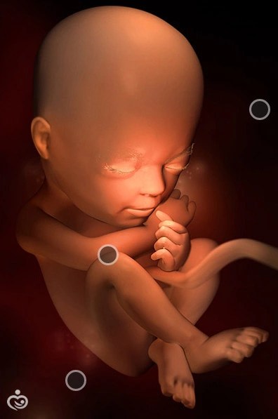
Billions of brain cells will develop over the next few weeks. Their job is to control all of your baby's movements, senses, and basic life functions like breathing.
The lungs begin to produce a substance that allows the lungs to swell and fill with air after birth, and the fetus begins to “breathe”. The frequency of respiratory movements is 50-60 per minute.
The height of the bottom of the uterus is 4 cm above the navel. The uterus grows, which can cause discomfort in the spine, joints, so a special bandage may be required.
24th week of pregnancy

The baby is still small, his weight is 600 g, and his height is about 33 cm. The child actively responds to the appeal to him. The inner ear is already fully formed (vestibular apparatus), he began to understand where the top is, and where the bottom is, the movements in the uterine cavity become more meaningful.
Mom adds about 500 grams per week. There may be swelling of the legs, so it is important to choose comfortable shoes, to give rest to the legs.
25th week of pregnancy

Fetal growth - 30-32 cm, weight 750 grams. Meconium is formed in the large intestine - the baby's first stool, which will completely pass within a few days after birth. The musculoskeletal system is actively developing, bones are being strengthened.
Mom may feel signs of anemia (anemia), which develops due to iron deficiency. Fatigue, pallor, fatigue and tachycardia are a reason to consult a therapist and take blood tests for anemia.
26th week of pregnancy

Height 34 cm, weight 900 grams.
There is an active development of the lungs, they are filled with a special substance that will not allow the lungs to stick together after the first breath.
The child clearly distinguishes periods of sleep and wakefulness. Mom feels his activity in the movement in her stomach. If you are lucky, then the periods of sleep and activity for you and your child will coincide.
27th week of pregnancy

The body weight of the fetus is already about a kilogram, and the height is 34 cm. Growth hormone begins to be produced in the pituitary gland. And in the thyroid gland - hormones that regulate metabolism.
In connection with involuntary contractions of the diaphragm, the mother may feel the hiccup-like movements of the baby. In adults, such movements are accompanied by the closure of the vocal cords, therefore, a characteristic “hiccup” sound occurs, and in a baby, this space is filled with liquid before childbirth, so such a “hiccup” is silent.
There may be new sensations in the legs - tingling, goosebumps or even cramps. This is an occasion to consult a doctor for further examination and treatment.
28th week of pregnancy
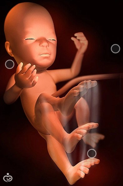
Now your baby is starting to close and open his eyes, which until now have not completely closed. The iris of the eyes has acquired color due to pigment, although this coloration is not final. Up to a year in children, eye color can change.
At week 28, in the case of multiple pregnancy, the mother receives a "sick leave". Weight gain by this time is 7-9 kg. At this time, Rh-negative mothers are given immunoglobulin.
29th week of pregnancy
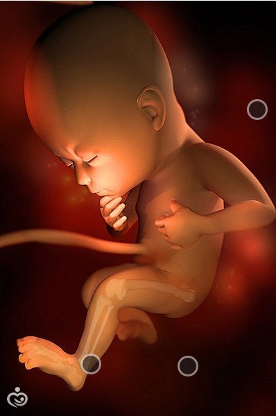
The child has a length of 36-37 cm, weighs approximately 1300 grams and is becoming stronger and more active. We can say that he also shows character. The child differently the child reacts to different products, sounds, light.
A woman suffers from heartburn, heaviness after eating. There may be frequent urination or even false urges.
30th pregnancy

In the coming weeks, your baby will actively gain weight. Adipose tissue will carry out the function of thermoregulation after birth, provide energy, protect organs. The movements of the baby will become less active, which is associated with an increase in its size. But if you do not feel any shocks in the usual mode, be sure to tell your doctor. Pregnant women may feel breast swelling and notice the release of colostrum.
At this time, a certificate of incapacity for work is issued for a singleton pregnancy.
From the 28-30th week of pregnancy, regular CTG (cardiotocography) begins to assess the condition of the fetus. With CTG, the fetal heartbeat, uterine tone and motor activity are evaluated.
31st week of pregnancy

Before birth, the little man will be in the fetal position, because otherwise he will no longer fit in the uterine cavity, his weight is 1600 g, and his height is already 40 cm.
These days, an important event occurs in male fetuses - testicles on their way to the scrotum. In girls, the clitoris is practically formed.
Weekly weight gain - 300-400 grams. At these times, edema may appear and pressure may increase, which may be a symptom of a formidable complication - preeclampsia. Therefore, the expectant mother should be as attentive as possible to any changes in well-being.
At a period of 30-32 weeks, an ultrasound of the third trimester is performed with dopplerometry - an assessment of blood flow.
32nd week of pregnancy

This is an important week, another critical deadline has passed. Babies born at this time are healthy and fully functional. By this week, all major organs are fully functional, except for the lungs, which need a little more time to fully mature.
The expectant mother may experience pain in the joints, pubic joint. Wearing a bandage, swimming can alleviate these phenomena.
33rd week of pregnancy
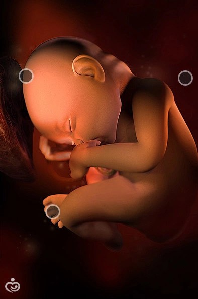
It’s getting harder for the baby to move, he is already 44 cm and weighs about 2 kg. Many babies like to live in the rhythm of mom: sounds, food and walks can affect the activity of the child.
The height of the bottom of the uterus is 34 cm from the level of the pubis. It becomes more difficult for mom to walk or exercise a lot, a break is required.
34th week of pregnancy

In a few weeks, your boy or girls begin to prepare to meet their parents. The original white grease that covers the skin begins to thicken, accumulating in the axillary and inguinal folds, behind the ears. At the time of the first toilet of the newborn, the lubricant will be removed. Height 47 cm, weight 2200-2300 g.
Pregnant women begin to feel false contractions - preparing the muscles for the birth process.
35th week of pregnancy

The fetus is preparing for birth, finally occupying the correct position, upside down. Approximately 97% of children are located in this way. The remaining 3% may be in a pelvic-down or even transverse position. Fetal height 47-48 cm, weight 2300-2500 g.
Almost all expectant mothers experience shortness of breath at this time.
36th week of pregnancy
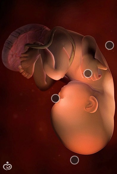
The baby continues to store fat, which is essential after birth for energy and warmth. The sucking muscles are ready to go: after birth, the baby will be hungry and will ask to be fed for the first time.
The height of the bottom of the uterus is 36 cm. The hormonal restructuring of the woman's body begins to prepare for childbirth - prostaglandins are also produced.
37th week of pregnancy
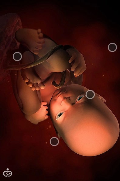
Finger movements become more coordinated, soon he will be able to grab your finger. The accumulation of subcutaneous fat continues, its volume is approximately 15% of the baby's weight. Height 48-49 cm, weight 2600-2800 g. The vellus hair covering the body gradually disappears.
The pregnant woman feels the harbingers of childbirth - the omission of the bottom of the uterus, a decrease in the volume of the abdomen, loosening of the stool, increased training contractions, discharge of the mucous plug.
38-40 weeks of pregnancy

In obstetrics textbooks, exactly 38 weeks was the term of a full-term pregnancy. If your baby is born right now, it will be a full-term pregnancy, and the baby will not have any of the risk factors associated with a premature birth. All further events are aimed at preparing for childbirth.
The discharge of the mucous plug should be distinguished from the leakage of amniotic fluid. If there is too much discharge, see your doctor for a special test.
In most cases, the baby's head sank into the small pelvis at 38-39 weeks, this is called cephalic presentation. If the child lies evenly with his head down, this is called a longitudinal position, if a little at an angle, then oblique. There is also the concept of the position of the fetus: I position means that the back is turned to the left uterine wall, and II - to the right. An ultrasound will tell you how the fetus is located.

At birth, doctors evaluate the child's condition according to several parameters: activity, muscle tone, heartbeat, breathing, skin color, reflex reactions. The more points, the healthier your baby was born.
Here we are at the end of our journey through 40 weeks of fetal development during pregnancy. The most important thing is to be attentive to yourself and the words of your doctor, prescribing tests and ultrasounds, paying attention to all the sensations and enjoying pregnancy and future motherhood.